Anatomy from the 3D printer: how digitalisation is helping medical students to understand the human body
In a unique project, UW/H researchers are working with students to create virtual and haptic 3D models of body donors - taking education to a new level.
")
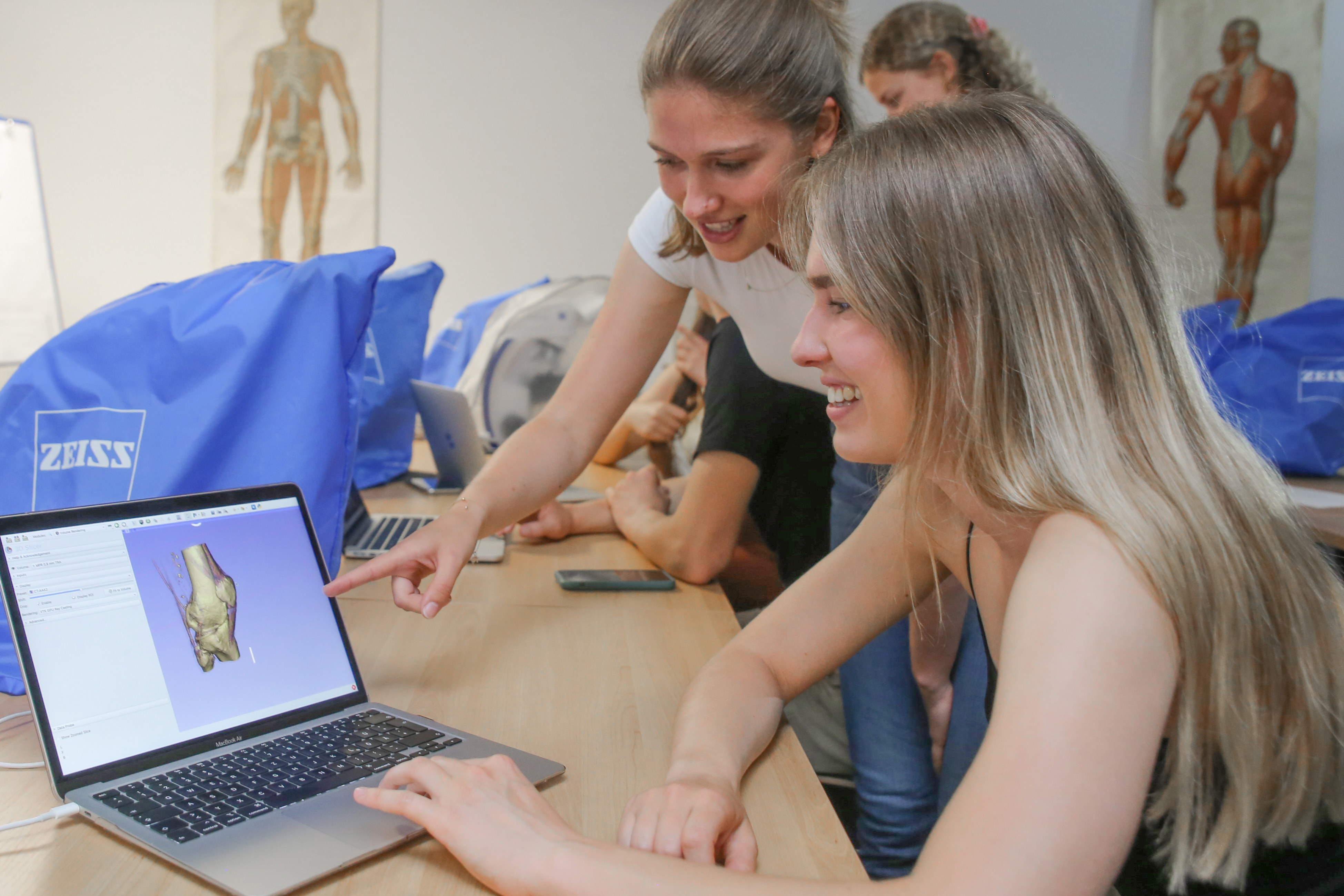
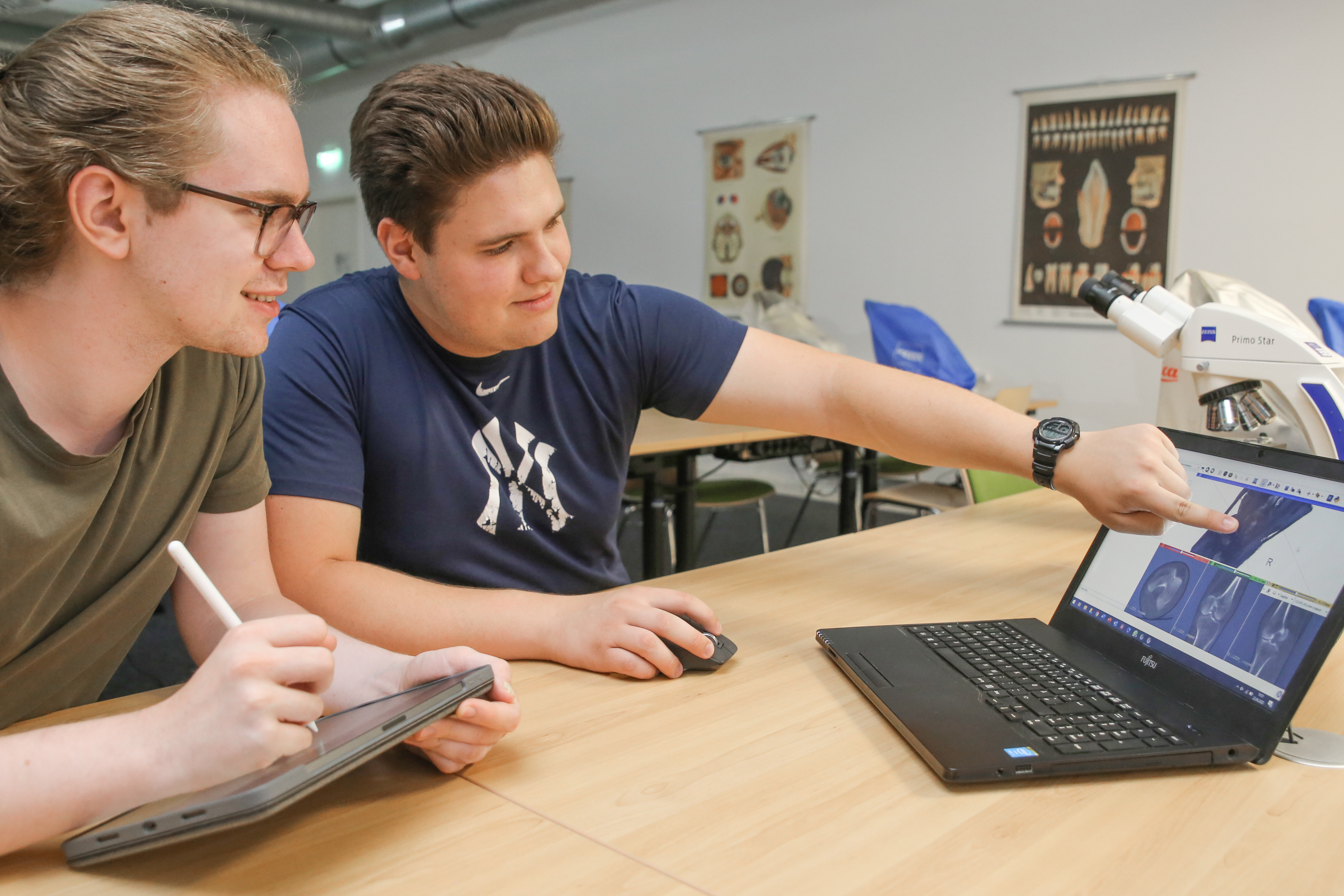
In order to familiarise themselves with human anatomy and prepare themselves as well as possible for their future appointment as a doctor, students of medicine at Witten/Herdecke University (UW/H) - as in many medical faculties worldwide - work on human body donors. As their number is limited, Dr Mona Eulitz (Chair and Institute of Anatomy and Clinical Morphology) and Priv.-Doz. Dr Christoph Stückle (Chair of Clinical Radiology, Prof. Dr Patrick Haage) have launched a special project at the UW/H: Two of the four body donors, which are available to each new year group for a period of two years, are scanned in thin layers in a modern computer tomography (CT) using X-rays. From the data obtained, the students independently create high-quality virtual 3D models of the bodies - both the soft tissue and the skeleton as well as the joints and their visible changes with age can be depicted in this way. This means that any number of students can learn simultaneously on their own 3D models on the screen and compare them later in the dissecting room with the original structures on which they are based.
"The fact that the students scan the body donors also offers the advantage that an 'incorrect dissection', in which a muscle or a large nerve was severed too early, for example, is no longer as serious. Finally, we can examine the entire body virtually at any time in its original state," says Dr Christoph Stückle.
Virtual models become tangible using 3D printers
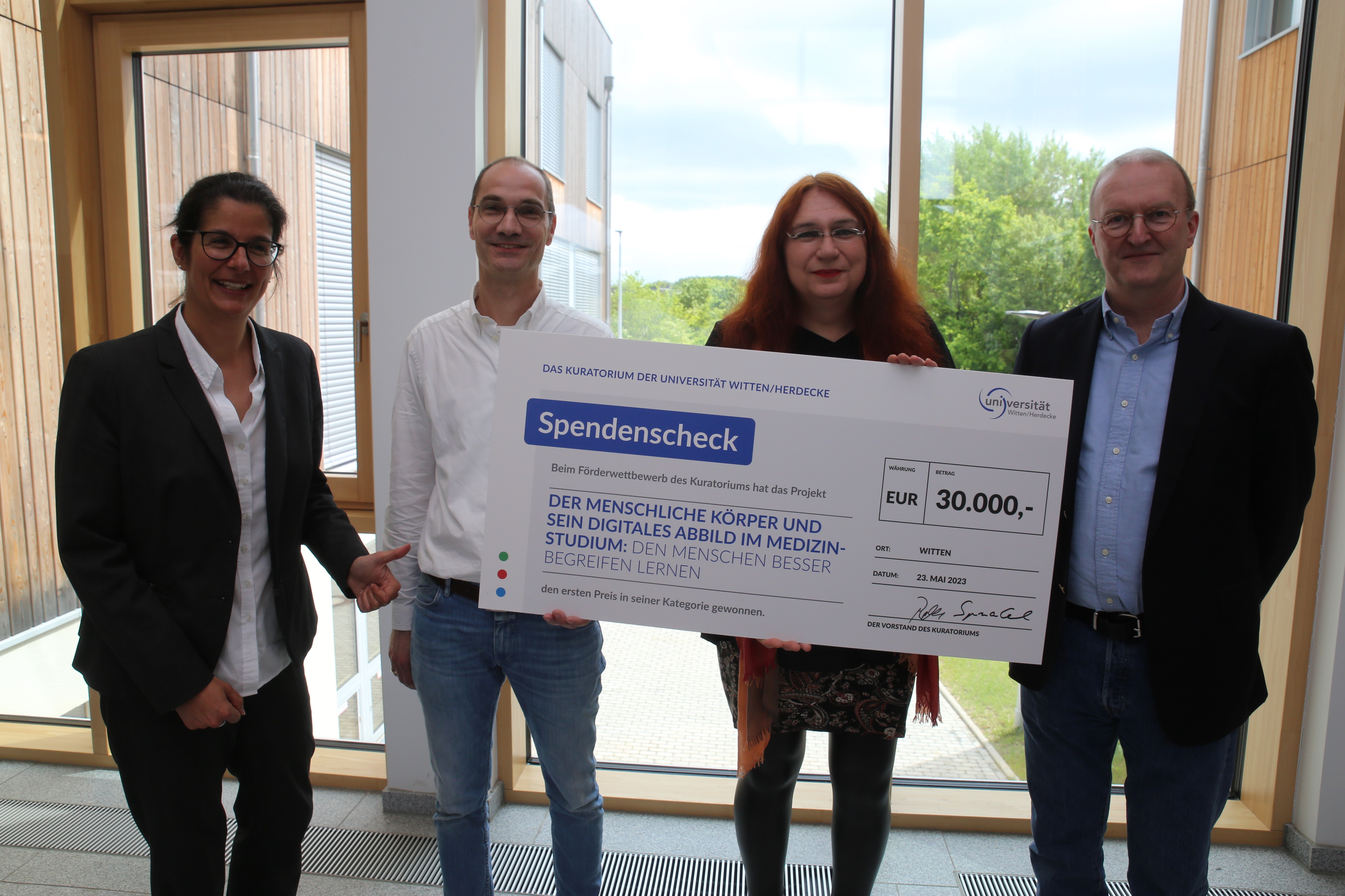
A flagship project that the researchers will be expanding over the next two years thanks to funding of 30,000 euros from the Board of Trustees of the UW/H. The students' virtual 3D models are to be printed in a modern 3D printer that can be purchased with the funds. This will allow the budding doctors to hold the models in their hands and examine them directly; the authentic replicas of real bodies will help them to understand anatomy much more easily. "Commercial models of idealised standard bodies are not even capable of reproducing this diversity," says Dr Eulitz. "This means that the project not only leads to greater learning success, but in the best-case scenario also to an increased interest in anatomy, radiology and pathoanatomy," adds Dr Stückle. In addition, the models will be transferred to a virtual reality environment so that students can take a virtual journey through their body donor and thus train and improve their spatial orientation in the human body.
Excellent preparation for later professional life
The continuously growing number of CT scans and 3D models also makes it possible to build up a unique collection of anatomical variations and pathological changes. "In future, we also want to carry out several scans per body donor with different types of radiation," says Dr Stückle. Thanks to the funding, the researchers will also be able to purchase an optical 3D scanner. This scanner digitises internal organs, some of which are more difficult to differentiate using CT.
Last but not least, practical skills are trained through the project: Computed tomography is one of the most frequently used imaging procedures in the everyday work of doctors. Through the intensive use of the procedure, UW/H students learn about the possibilities and limitations of CTs and can link them to the corresponding anatomical structures. "By subsequently creating their own virtual and, in future, haptic 3D models from the CT data, the students at our university are definitely among the pioneers in medical training," says Dr Eulitz.
Further information: The Board of Trustees of the UW/H is also funding two other projects with 10,000 and 20,000 euros: The student initiative "Medical Students for Choice Witten" wants to use the money to contribute to holistic education and destigmatisation of abortions. To this end, it organises so-called papaya workshops, among other things. The Chair of Pharmacology and Toxicology is using the funding to research new antibiotically effective substances. Further information is available on the UW/H's Instagram and LinkedIn channels.
Photos for download
 and Dr Mona Eulitz (3rd from left) receive funding from the UW/H Board of Trustees for their project. (Photo: UW/H)")
For their project, Priv.-Doz. Dr Christoph Stückle (2nd from left) and Dr Mona Eulitz (3rd from left) receive funding from the UW/H Board of Trustees for their project. (Photo: UW/H)
")
")
")
")
Contact person
")
Svenja Kurth , M. A.
Team Leader Communication
Administration | Communication & Marketing
Alfred-Herrhausen-Straße 48
58455 Witten
Room number: 2.F05 | 2.028