Making the invisible visible in the human body.
Prof. Dr Maximilian Ackermann, pathologist at the Helios University Hospital Wuppertal of Witten/Herdecke University, is honoured with the Lennart Nilsson Prize of the Karolinska Institute for his scientific photography.
")
How do blood vessels in the brain change in Alzheimer's disease? What happens in the lungs during a coronavirus infection? And how does cancer tissue spread? Prof Dr Maximilian Ackermann provides answers to these questions with impressively precise and artistic images from inside the human body. For his research, the pathologist and anatomist from Helios University Hospital Wuppertal at Witten/Herdecke University, RWTH Aachen University Hospital and the associated independent institute of anatomy at Mainz University Medical Centre has been awarded this year's Lennart Nilsson Prize by the Karolinska Institute in Stockholm - one of the world's most prestigious awards for scientific photography.
The prize honours Prof. Ackermann's pioneering work in this field, which has made a decisive contribution to improving our understanding of the progression of cancer, Alzheimer's and lung diseases. The latest high-resolution technologies such as hierarchical phase contrast tomography (HiP-CT) and scanning electron microscopy, which go far beyond the possibilities of conventional clinical imaging methods, are used for this purpose. With their help, the finest structures and vessels as well as pathological changes in organs can be visualised in three dimensions.
"The award is a great honour for me," says prizewinner Prof. Ackermann. "It spurs me on, like Lennart Nilsson, to go on a journey of discovery in the human body to make the invisible visible."
The prize, which is endowed with 120,000 Swedish kronor, is awarded annually and honours the photographer Lennart Nilsson, who achieved world fame with his scientifically fascinating yet aesthetic images of the inside of the body.
Further information:Prof Dr Maximilian Ackermann's academic work bridges the gap between basic research and its practical application in patient care. He focusses on the formation of new blood vessels and how these are influenced by medication or other therapies in cardiovascular diseases, cancer or wound and tissue repair. He has already been honoured with the prestigious Rudolf Virchow Prize and the Boehringer Ingelheim Prize for his research. Insights into his scientific photography and further background information on his work can be found here.
Picture credits:
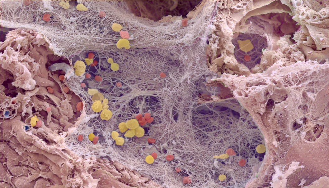
- The coloured scanning electron micrograph of a human lung with COVID-19 infection shows numerous alveoli with inflammatory cells (yellow), haemorrhages (red) and hyaline membranes (blue). Photo: Maximilian Ackermann.
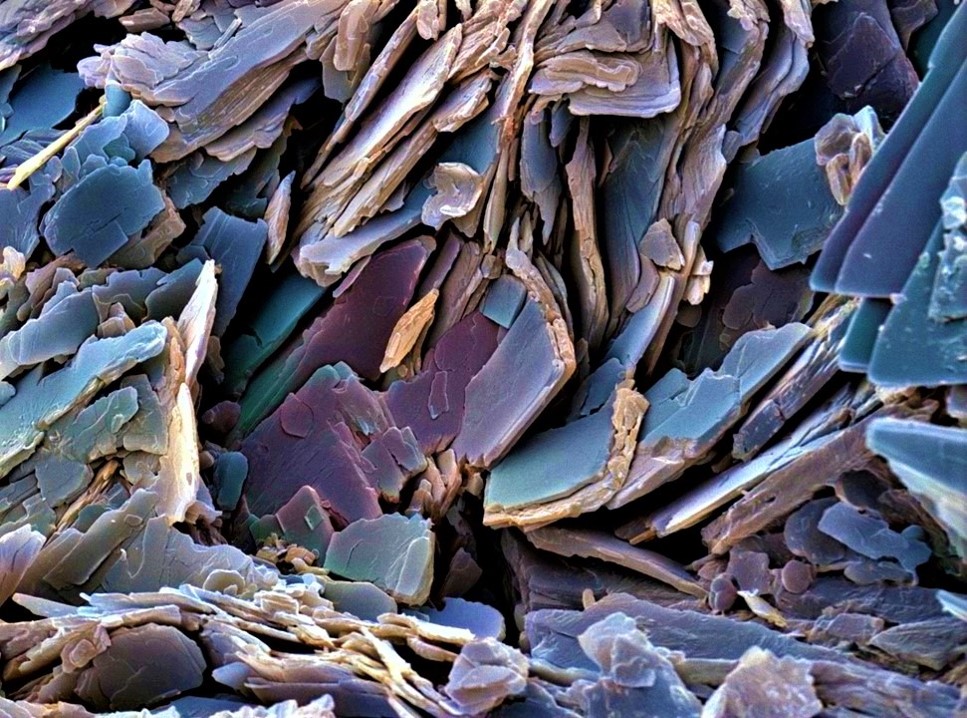
- The coloured scanning electron micrograph of a human gallstone resembles grey slate in its appearance. Photo: Maximilian Ackermann.
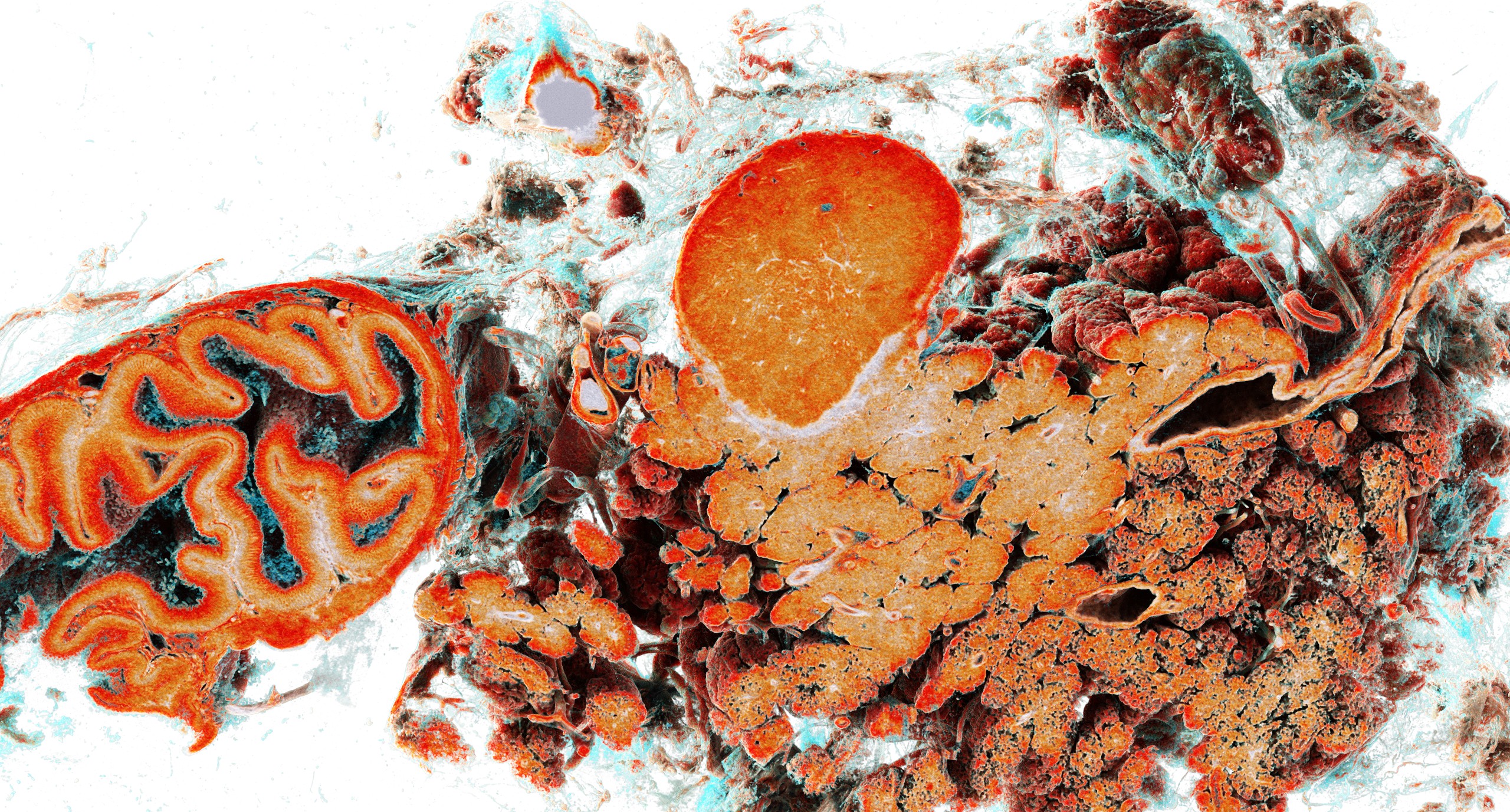
- Synchrotron-based HiP-CT scans of a Whipple resection specimen (duodenum and pancreas) with a 1.1 cm neuroendocrine tumour of the pancreatic tissue. The mucosa of the stomach and duodenum can be seen first, followed by the course of the intrapancreatic bile duct and the exocrine pancreas. An encapsulated, well-vascularised neuroendocrine tumour can be seen in the pancreas. Photo: Maximilian Ackermann.
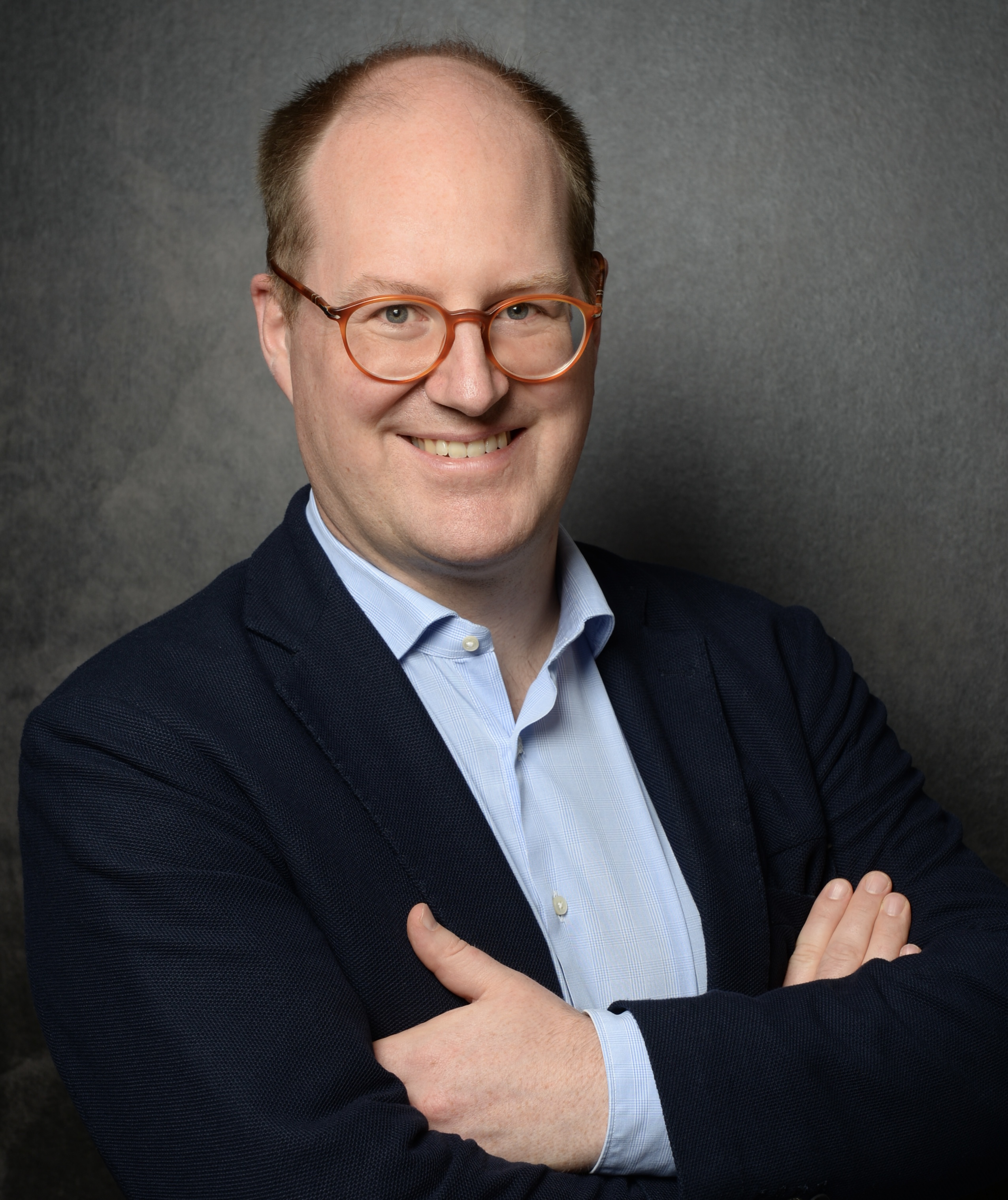
- Prize winner Prof. Dr Maximilian Ackermann. Photo: Maximilian Ackermann
Photos for download
")
. (Photo: Maximilian Ackermann)")
")
")
Contact person
")
Miriam Kreimeyer
Communications Officer
Administration | Communication & Marketing
Alfred-Herrhausen-Straße 48
58455 Witten
Room number: 2.F05