New X-ray technology shows vascular damage in intact COVID-19 lungs for the first time
Research team confirms changes in blood vessels due to infection with SARS-CoV-2
")
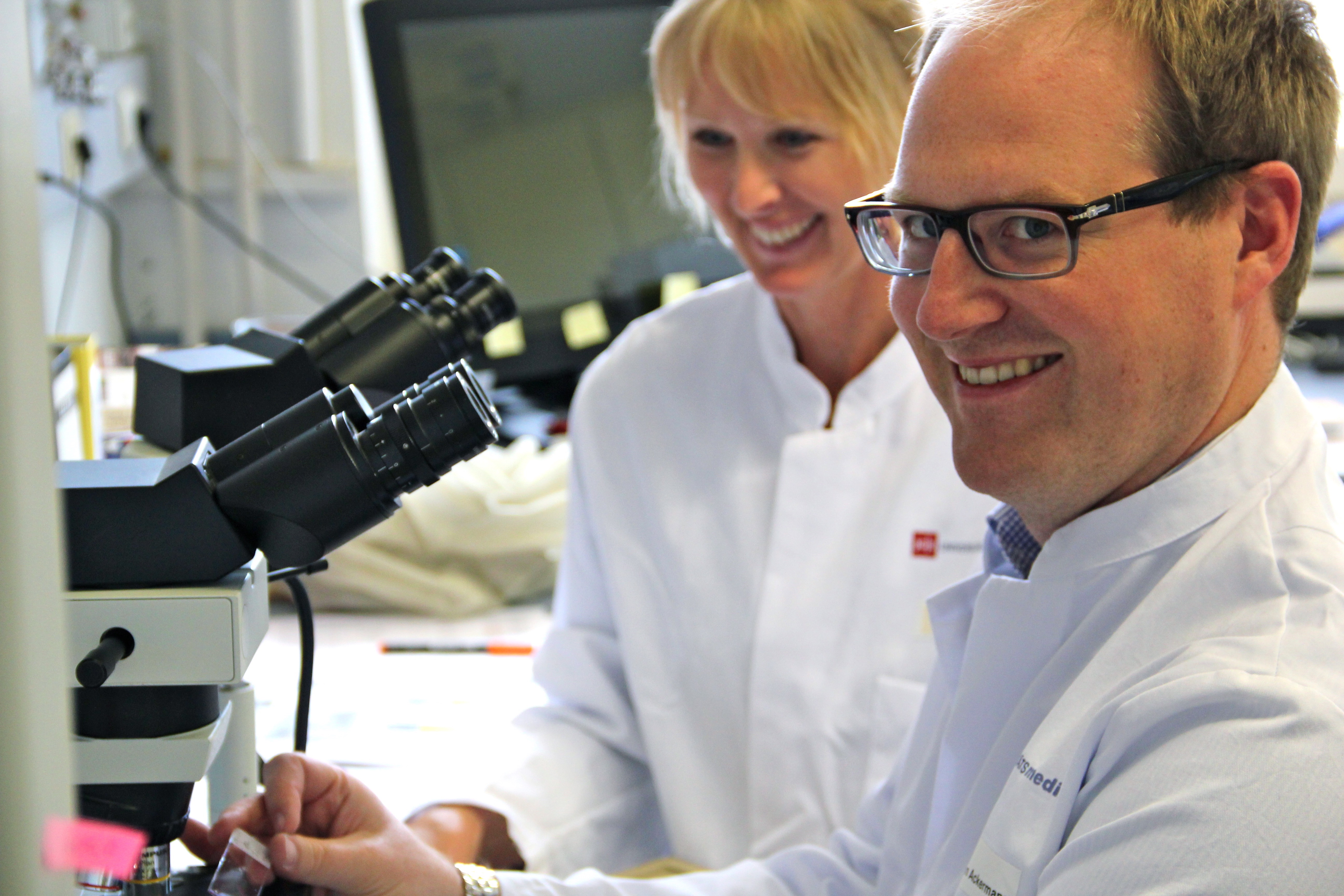
If the SARS-CoV-2 coronavirus enters the lungs, it causes massive tissue damage. A characteristic consequence of the infection is, among other things, blockage of the pulmonary vessels due to localised excessive blood clotting. An international research team led by PD Dr Maximilian Ackermann from the Institute of Pathology and Molecular Pathology at Helios University Hospital Wuppertal at Witten/Herdecke University and Prof. Dr Danny Jonigk from Hannover Medical School has now been able to demonstrate for the first time using a highly innovative non-destructive X-ray technique that severe COVID-19 causes massive remodelling of the finest blood vessels, with normally separate blood systems connecting with each other unusually frequently.
The researchers examined the lungs of COVID-19 victims in collaboration with the European Synchrotron Research Facility (ESRF), the world's third-largest particle accelerator in Grenoble, France. Thanks to the latest technology, a three-dimensional image of the entire organ was produced for the first time using high-resolution X-rays. The work has resulted in two publications in renowned scientific journals: The technical procedure has been published in "Nature Methods", the clinical application in the "American Journal of Respiratory and Critical Care Medicine (Blue Journal)".
HiP-CT shows an entire organ in three dimensions without damaging it
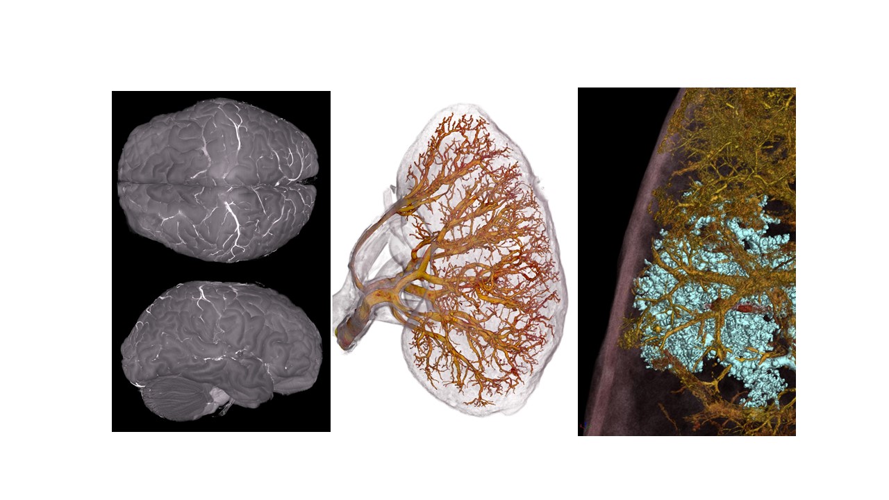
The new X-ray technology works in a similar way to computer tomography (CT) in hospitals. However, the resolution is a hundred times higher. "In clinical CT scans, we can only show blood vessels in the millimetre range," explains vascular specialist PD Dr Ackermann. The new technology called Hierarchical Phase Contrast Tomography (HiP-CT) is able to image the finest vessels with a diameter of less than five micrometres - this corresponds to around a tenth of the thickness of a hair. HiP-CT makes it possible to penetrate deep into the organs and visualise even the smallest structures down to individual cells. "This resolution was previously only possible with a microscope, but only in two dimensions and for small tissue samples," adds lung pathologist Prof Jonigk.
The new HiP-CT method can significantly exceed this resolution. The new technique makes it possible for the first time to image an entire organ in three dimensions and at a high magnification without damaging it. "This allowed us to examine structures that are at the limits of resolution and gain an overview of the changes in the entire organ system," emphasise both scientists. The Extremely Brilliant Source at the European research centre ESRF was created in an international collaboration between 22 countries, including the USA and Japan, with an investment volume of more than 150 million euros. He is all the more pleased that the Wuppertal-based company is one of the first users of the new technology. "When all major research facilities were in lockdown at the beginning of the COVID-19 pandemic, we were able to work with our colleagues in Grenoble to convince the French government to ramp up the research reactor in Grenoble for our COVID-19 research," Ackermann continues. "It certainly helped that we were able to show last spring that COVID-19 is not primarily a purely viral respiratory disease, but rather a systemic vascular disease that we wanted to characterise in more detail."
COVID-19 leads to "short circuits" in the blood vessels of the lungs
Using the new X-ray technology, the scientists have now been able to show how the vascular system changes in COVID-19. There are two separate blood systems in the lungs - one is part of the pulmonary circulation and is responsible for supplying oxygen to the entire body, while the other supplies the lung tissue itself with the vital gas directly from the aorta. In a healthy lung, there are sometimes a few connections between small vessels of the two systems.
In the damaged COVID-19 lung, however, the two blood systems formed numerous such connections in many areas. "This large number of vascular short circuits functions like a wide-open floodgate; they ensure that the oxygen supply to the entire body no longer functions via many thrombi," explains first author PD Dr Ackermann. "Due to the many short-circuit reactions, the lungs manage to compensate for the lack of oxygen caused by the SARS-CoV-2 infection in the short term and new blood vessels are even formed. However, these newly formed blood vessels can only heal the considerable vascular damage in vain," adds PD Dr Ackermann.
The two scientists are certain that the networking of pathology and radiology through this brilliant, high-resolution technology will revolutionise medical imaging and our understanding of the structure of our body. "The new HiP-CT X-ray technology not only allows us to resolve an organ in three dimensions at high resolution, but also gives us the opportunity to characterise pathological regions at a molecular level afterwards," adds Anatomist Ackermann. As part of the "Human Organ Project", the research team has already started to create a more extensive organ atlas. In addition to the COVID-19-damaged lung, it already contains images of several healthy human organs such as the brain, lungs, heart, kidneys and spleen from deceased body donations. The international team is convinced that HiP-CT X-ray technology will also provide new insights into numerous diseases, including cancer and Alzheimer's.
The work was carried out in cooperation between the Institute of Pathology and Molecular Pathology at the Helios University Hospital Wuppertal of Witten/Herdecke University and, among others, the Institute of Pathology at Hannover Medical School, the German Centre for Lung Research, the University Medical Centre Mainz and University College London.
Original publications:
- Walsh C, Tafforeau P, Wagner WL, Jafree DJ, Bellier A, Werlein C, Kühnel MP, Boller E, Walker-Samuel S, Robertus JL, Long DA, Jacob J, Marussi S, Brown E, Holroyd N, Jonigk DD, Ackermann M, Lee PD. Multiscale three-dimensional imaging of intact human organs down to the cellular scale using hierarchical phase-contrast tomography. Nature Methods, November 4, 2021.
https://www.nature.com/articles/s41592-021-01317-x
DOI: 10.1038/s41592-021-01317-x - Ackermann M, Tafforeau P, Wagner WL, Walsh C, Werlein C, Kühnel MP, Länger FP, Disney C, Bodey A, Bellier A, Verleden SE, Lee PD, Mentzer SJ, Jonigk DD. The bronchial circulation in COVID-19 pneumonia. American Journal of Respiratory and Critical Care Medicine, November 4, 2021.
https://www.atsjournals.org/doi/abs/rccm.202103-0594IM
DOI: 10.1164/rccm.202103-0594IM
Photos for download
")
. (Photo: ERSF)")
Contact person
")
Svenja Malessa
Press Officer
Administration | Communication & Marketing
Alfred-Herrhausen-Straße 48
58455 Witten
Room number: 2.F05